Klippel-Feil Syndrome
Synonyms and related keywords: low hairline,
short neck, cervical spine disorder, synkinesia, Klippel Feil syndrome
INTRODUCTION
In
1912, Maurice Klippel and Andre Feil were independently the first to describe
Klippel-Feil syndrome. They described patients who had a short neck, increased
range of motion (ROM) in the cervical spine, and a low hairline. Feil
subsequently classified the syndrome into 3 categories. Type I is described as
a massive fusion of the cervical spine. Type II is present when the fusion of 1
or 2 vertebrae occurs. Type III occurs when thoracic and lumbar spine anomalies
are associated with type I or type II Klippel-Feil syndrome.
Patients
with Klippel-Feil syndrome usually present during childhood but may present
later in life. The challenge to the clinician is to recognize the associated
anomalies that can occur with Klippel-Feil syndrome and to perform the
appropriate workup for diagnosis.
Frequency: The true incidence of Klippel-Feil syndrome is
unknown. No one has ever studied a cross-section of healthy people to determine
the true incidence.
The
incidence of Klippel-Feil syndrome has been investigated in 2 studies, using 2
different means. Gjorup reviewed all of the radiographic cervical spine films
in a single hospital in Copenhagen.
From these radiographs, he determined an incidence of 0.2 cases per 1000
people. Brown reviewed 1400 skeletons from the Terry collection at Washington
University School of Medicine. He found an incidence of 0.71%.
Etiology: The etiology of Klippel-Feil syndrome and its
associated conditions is unknown. The syndrome can present with a variety of
other clinical syndromes, including fetal alcohol syndrome, Goldenhar syndrome,
and other anomalies in the extremities. Gunderson suggested that it is a
genetic condition, while Gray found a low incidence of inheritance. Others have
considered it to be some type of global fetal insult, which could explain the
other associated conditions. Others have considered it to be caused by vascular
disruption. The true etiology has yet to be determined.
Clinical: Clinical presentation is varied because of all of
the associated syndromes and anomalies that can occur in patients with
Klippel-Feil syndrome. A complete history and careful physical examination may
reveal some associated anomalies. From an orthopedic standpoint, most of the
workup is with imaging (see Imaging Studies).
The condition is detected throughout life, often as
an incidental finding. Patients with upper cervical spine involvement tend to
present at an earlier age than those whose involvement is lower in the cervical
spine. Most patients present with a short neck, decreased cervical ROM, and a
low hairline, which occurs in 40-50% of patients. Decreased ROM is the most
frequent clinical finding. Rotational loss usually is more pronounced than is
the loss of flexion and extension.
Other patients present because of cosmesis or
facial asymmetry. Neurological problems may develop in 20% of patients.
Pouliken found that 5 of 19 patients with Klippel-Feil syndrome had
neurological involvement; of these 5 patients, 2 had neurological problems due
to hypermobility at one level. Occipitocervical abnormalities were the most
common cause of neurological problems (see Images 1-4). Some patients present with pain.
Hensinger and colleagues described the
constellation of anomalies that can occur with Klippel-Feil syndrome, and
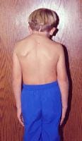
others have added to this list. Scoliosis occurs in approximately 60% of
patients (see Images 5-6). In some patients, this is
congenital scoliosis (see Image 7) due to the involvement of other parts
of the thoracic or lumbar spine. Other patients develop compensatory scoliosis
in the thoracic spine to compensate for the cervical or cervicothoracic
scoliosis. In addition to the fusion anomalies in the cervical spine, cervical
spinal stenosis can occur. While uncommon, this can increase the risk of
neurological involvement.
Anomalies of the craniocervical junction can cause
instability at lower segments. Traumatic tetraplegia has been reported
following minor trauma. A Sprengel anomaly occurs in 20-30% of patients (see Image 7). Always check ROM of the shoulders.
Look for an omovertebral bone (see Image 8). This is an osteocartilaginous
connection that tethers the scapula to the spine. An omovertebral bone ossifies
with age, further limiting the ROM.
A CT
scan best demonstrates an omovertebral bone; however, palpation or radiographs
also can detect an omovertebral bone. Other upper extremity anomalies occur
less frequently. A thorough examination of the ROM and function of the upper
extremity must be performed.
Synkinesia is mirror movement of the upper
extremity. Patients with this condition are unable to perform a movement of the
right hand without performing the same movement of the left hand (see Image 9). This is disabling in activities of
daily living (ADL). Synkinesia often may be improved with therapy and usually
improves with age.
Renal anomalies are common in individuals with
Klippel-Feil syndrome, and they can be quite serious. Hensinger reviewed 50
patients, and 41 of them had an intravenous pyelogram (IVP). Renal anomalies
were present in 16 (34%). Minor renal anomalies were detected in 6 individuals,
including a double collecting system, renal ectopia, and bilateral tubular
ectasia. Major renal anomalies were detected in 10 individuals, including
hydronephrosis, absence of a kidney (see Image 10), and a horseshoe kidney. As Hensinger
notes, the patient of Klippel and Feil died of renal failure and uremia.
Patients with Klippel-Feil syndrome now have ultrasound as the initial test to
demonstrate the presence of 2 functioning kidneys.
Torticollis and facial asymmetry occur in 21-50% of
patients with Klippel-Feil syndrome. These patients also may have a muscular
torticollis. Craniofacial anomalies also can occur.
Hearing loss is common with Klippel-Feil syndrome.
The hearing loss can be sensorineural, conductive (one third of cases), or
mixed. Hensinger found the incidence of hearing loss to be 36%. Early
audiometric evaluation and otologic evaluation are indicated in all children
when the diagnosis of Klippel-Feil syndrome is established.
Cardiovascular anomalies occur in 14-29% of cases.
The most common cardiovascular defect is an interventricular septal defect.
Other less common anomalies are congenital limb deficiencies, craniosynostosis,
ear abnormalities, iniencephaly, and craniofacial abnormalities.
INDICATIONS
Patients with Klippel-Feil syndrome present at
different ages with varying clinical presentations. Indications for the
complexity of the workup vary individually. For the orthopedic surgeon, the
most usual indications for surgery depend upon the amount of deformity, its
location, and its progression with time. Other indications include instability
of the cervical spine and/or neurological problems. These indications can occur
with craniocervical junction anomalies and also when 2 fused segments are
separated by a normal segment.
Some patients present early in life with complex cervical and cervicothoracic deformity that is progressive and disfiguring. Some of these patients require cervical spine fusions to prevent progression.
Some patients present early in life with complex cervical and cervicothoracic deformity that is progressive and disfiguring. Some of these patients require cervical spine fusions to prevent progression.
Other patients may develop compensatory or
associated congenital scoliosis, which also can be progressive over time and
requires fusion to prevent progressive deformity. Over one half of the patients
in Hensinger's study had scoliosis. Treatment of the scoliosis with bracing or
surgery was required in 18 of the 50 patients.
RELEVANT ANATOMY AND CONTRAINDICATIONS
WORKUP
- Plain radiography is the basis for the diagnosis of Klippel-Feil syndrome.
- Initial studies include anteroposterior (AP) and lateral views of the cervical spine.
- If anomalies are found, carefully assess the craniocervical junction to detect anomalies at that level.
- Flexion-extension radiographs are indicated if instability is suspected at the craniocervical junction or if 2 fused segments are separated by an open segment.
- Obtain plain radiographs of the entire spine to detect other spinal anomalies.
- Examine the chest to rule out involvement of the heart. Examine the chest wall for the possibility of rib anomalies, which can include multiple rib fusions. Rib fusions can be revealed with plain radiography.
- CT scan often is more useful at the spinal level.
- For patients being evaluated for surgery, CT scan with 3-dimensional reconstruction can be a very valuable tool to assess the anatomy.
- A unilateral unsegmented bar or cervical stenosis may be revealed on a CT scan, which helps the physician plan the surgical procedure.
- MRI
- MRI is indicated in patients with neurological deficits.
- Flexion-extension MRI may reveal cord compression and is useful in evaluating spinal stenosis.
- In patients with neurological deficits, obtain an MRI of the entire spine to search for central nervous system anomalies, such as a syringomyelia.
- Ultrasound initially is performed to visualize the kidneys.
- Perform intravenous pyelography if any kidney abnormality is suspected with ultrasound.
Other Tests:
- Due to the high incidence of hearing loss with Klippel-Feil syndrome, an audiologist or an otologist should evaluate all children.
TREATMENT
Medical therapy: Medical therapy is dependent upon the congenital
anomalies present in the syndrome. Primary care physicians may not be familiar
with all of the possible associated anomalies. Patients with genitourinary
abnormalities are referred to a nephrologist or urologist. Patients with
cardiovascular abnormalities are cared for by a cardiologist or primary care physician.
Patients with auditory abnormalities are referred to an audiologist or
otologist.
Surgical therapy: Surgery is indicated for a variety of situations in
patients with Klippel-Feil syndrome. Due to fusion anomalies and the difference
in growth potential between the 2 sides of the spine, deformity may be
progressive. Instability of the cervical spine can develop because of
craniocervical abnormalities. Instability of the cervical spine also can
develop between 2 sets of fusion anomalies separated by normal segment.
Neurologic deficits or persistent pain are indications for surgery. Development
of a compensatory curve in the thoracic spine may require surgical intervention
or bracing. Symptomatic spinal stenosis may require decompression and fusion.
Koop
and colleagues studied 13 cases of children who were skeletally immature with a
variety of disorders that caused instability of the upper part of the cervical
spine from the occiput to C5. These researchers were looking at the efficacy of
posterior arthrodesis and halo-cast mobilization. Many of these patients did
not have Klippel-Feil syndrome, but the surgical indications were instability.
Posterior arthrodesis with external mobilization by halo-cast was carried out.
In 2 of the patients, internal fixation with wire was utilized. A solid
arthrodesis was obtained in 12 patients treated with autogenous bone graft. One
patient treated with allograft rib developed a pseudoarthrosis.
Koop
et al cautioned that the use of wires for fixation carries risk of neural
injury and often is not applicable in children with anomalous vertebra. They
stress the need for delicate exposure, decortication using an air drill, and
placement of autologous iliac graft. They recommended mobilization by
halo-cast, which they thought would minimize the risk of neural damage and
provide a reliable means of obtaining arthrodesis.
Preoperative details: Patients must have a comprehensive workup to detect
the anomalies previously mentioned. Adequate imaging studies must be obtained.
Three-dimensional CT scan reconstruction often is useful.
OUTCOME AND PROGNOSIS
FUTURE AND CONTROVERSIES
The true etiology and incidence of
Klippel-Feil syndrome is unknown. The syndrome can occur in association with a
wide variety of anomalies. Further studies may reveal the cause. The cervical
anomaly is a failure of segmentation that occurs in early fetal life. To
discover a cause and devise prevention or treatment is a challenge.


0 comments
Post a Comment